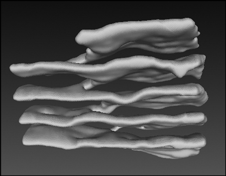
The endoplasmic reticulum (ER) is a continuous membrane system consisting of the nuclear envelope and a peripheral network of membrane tubules and sheets. ER sheets often form stacks, an arrangement that is likely required to accommodate a maximum number of membrane-bound polysome for secretory protein synthesis. How sheets are connected with one another was unknown until recently. As reported in Cell, the Rapoport Lab and their collaborators used a novel automated serial thin sectioning electron microscopy technique to analyze the 3D structure of stacked ER sheets of mouse neurons and of professional secretory cells of the salivary gland. The team discovered that ER sheets are connected by a novel membrane motif consisting of continuous twisted membrane surfaces with helical edges that have left or right-handedness. A theoretical model indicates that this configuration corresponds to a minimum of elastic energy of the sheet surface and its edges. The three dimensional structure resembles a parking garage and likely allows the optimal packing of ER sheets in the restricted volume of a cell.