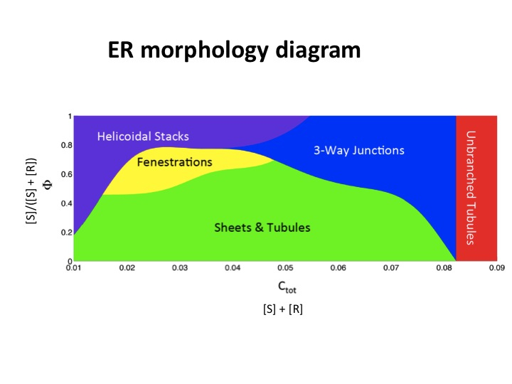
 The endoplasmic reticulum (ER) is an important membrane-bound organelle in all eukaryotic cells. Depending on cell type and functional state, the ER membrane can adopt different morphologies, including a network of interconnected tubules, and sheets that can contain fenestrations or be stacked on top of each other. How these different morphologies are generated is unclear. A collaboration of different groups, including the Rapoport group, resulted in a recent paper that presents a comprehensive theoretical model, which explains the formation and interconversion of virtually all known ER morphologies (Shemesh et al., 2014, Proc. Natl. Acad. Sci. USA PMID:25404289). The model is based on two types of membrane-shaping proteins (R- and S-type proteins), exemplified by the reticulons and lunapark, which both stabilize the high membrane curvature in cross-sections of tubules and sheet edges, but favor straight or concave sheet edges, respectively. Dependent on the concentrations of R- and S-type proteins, membrane morphologies can be generated that consist of tubules, sheets, sheet fenestrations, and sheet stacks with helicoidal connections. In a tubular ER network, lunapark stabilizes three-way junctions, i.e. small triangular sheets with concave edges. The model agrees with experimental observations and explains how curvature-stabilizing proteins determine ER morphology.