 In a recent article in Nature, the Pellman Lab reports that increased numbers of centrosomes can mimic and enhance the effects of a breast cancer oncogene. Centrosome amplification, a common cytoskeletal aberration in cancer cells, has long been recognized as a feature of human tumors, but its role in tumorigenesis has been unclear. This amplification is poorly tolerated in non-transformed cells; extra centrosomes are spontaneously lost in the absence of selection. Thus, the high frequency of centrosome amplification, particularly in more aggressive tumors, presents something of a paradox and raised the possibility that extra centrosomes could confer advantageous characteristics that promote tumor progression.
Using a 3D model system and other approaches to culture human mammary epithelial cells, the Pellman Lab found that centrosome amplification triggers cell invasion. This invasive behavior is similar to that induced by overexpression of the breast cancer oncogene ERBB2 and enhances invasiveness triggered by high-level expression of ERBB2. Their data indicate that, through increased nucleation of microtubules from centrosomes, centrosome amplification increases the activity of the small GTPase, Rac1, disrupting normal cell-cell adhesion and promoting invasion. Activation of Rac is a well-established feature of cancer cells and is thought to be critical for cancer cell invasion and metastasis. These findings demonstrate that centrosome amplification, a structural alteration of the cytoskeleton, can promote features of malignant transformation.
This work was in collaboration with the Thery Lab (Hospital Saint Louis), Brugge Lab (HMS, Cell Biology), and Polyak Lab (HMS & DFCI).
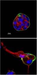
The image illustrates the ability of centrosome amplification to promote invasive behavior of mammary epithelial cells. Top image: normal, early stage organoids in 3-D cultures; bottom image: organoids from cells with centrosome amplification. Laminin V is in green, actin is in red, and DNA is in blue.